What are you looking for ?
Content on this site

Content on this site
contact: Sébastien BROT
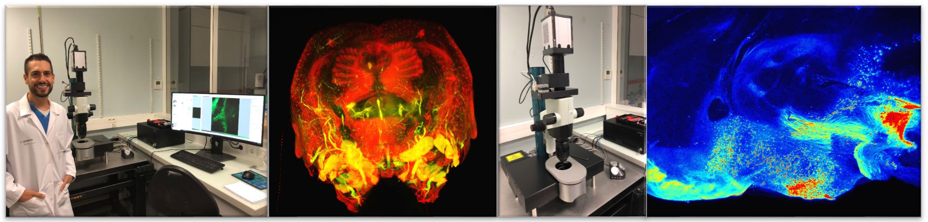
The ultramicroscope (LaVision Biotec) is a fluorescence microscope based on light-sheet illumination. The illumination is therefore perpendicular to the acquisition plane by a light sheet between 4 and 10 microns thick and the width of the entire sample.
In order to limit the artifacts the light sheet is divided into three sub-beams which converge towards the sample with different angles, which avoids the current shading phenomena with this technique. The microscope is equipped with a very sensitive camera (CMOS) and very resolutive (2560 × 2160 pixels per image). The available magnifications (from 1.26 to 12.6 X) allow an optimal exploration of whole organ up to cellular levels. It is equipped with 4 diode lasers: 488nm, 561 nm, 639nm and 785nm. This technology involves the use of clarified samples whose production techniques are now well established. The size of the volume that can be imaged is of the order of one cubic centimeter to a resolution of about one micrometer in the plane and three microns in depth. Booking via ImageUP
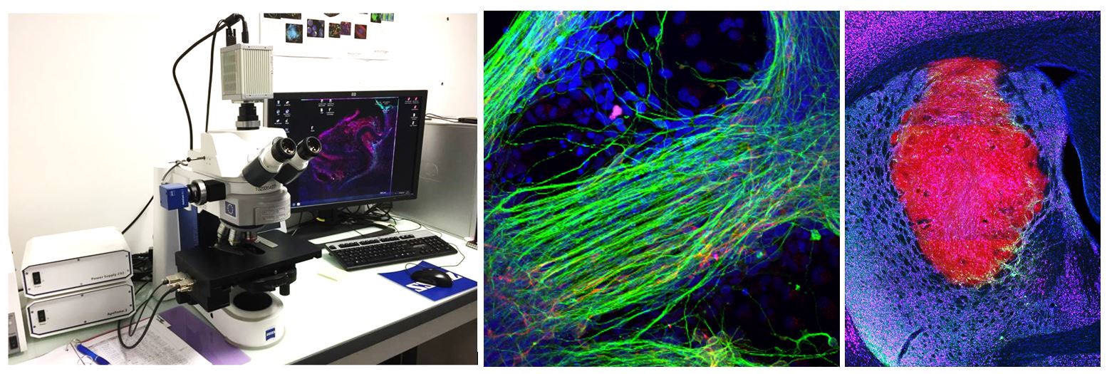
The Axio Imager 2 microscope is equipped with 6 filters for fluorescence, an ultra-sensitive Orca4 Flash camera from Hamamatsu, and the latest version of the ZEN software for acquisition. The microscope is equipped with an apotome module in order to be able to create optical sections of the fluorescent samples, free from scattered light. With structured illumination, only the focal plane appears in the image. A totally reliable method to prevent scattered light out of the focal plane, even in the thickest samples. The images obtained are strongly contrasted with the best possible resolution.
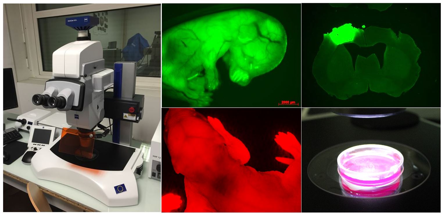
The Axio Zoom.V16 combines a 16x zoom with a high numerical aperture (NA> 0.25). This macroscope allows the observation of fluorescent samples (or not) at a macroscopic level (from a few cells to whole organisms). Images of very good quality can be obtained from specimens under blade / coverslip, but also through Petri dishes (eg embryos). This device allows you to acquire a rodent brain cut image in seconds.
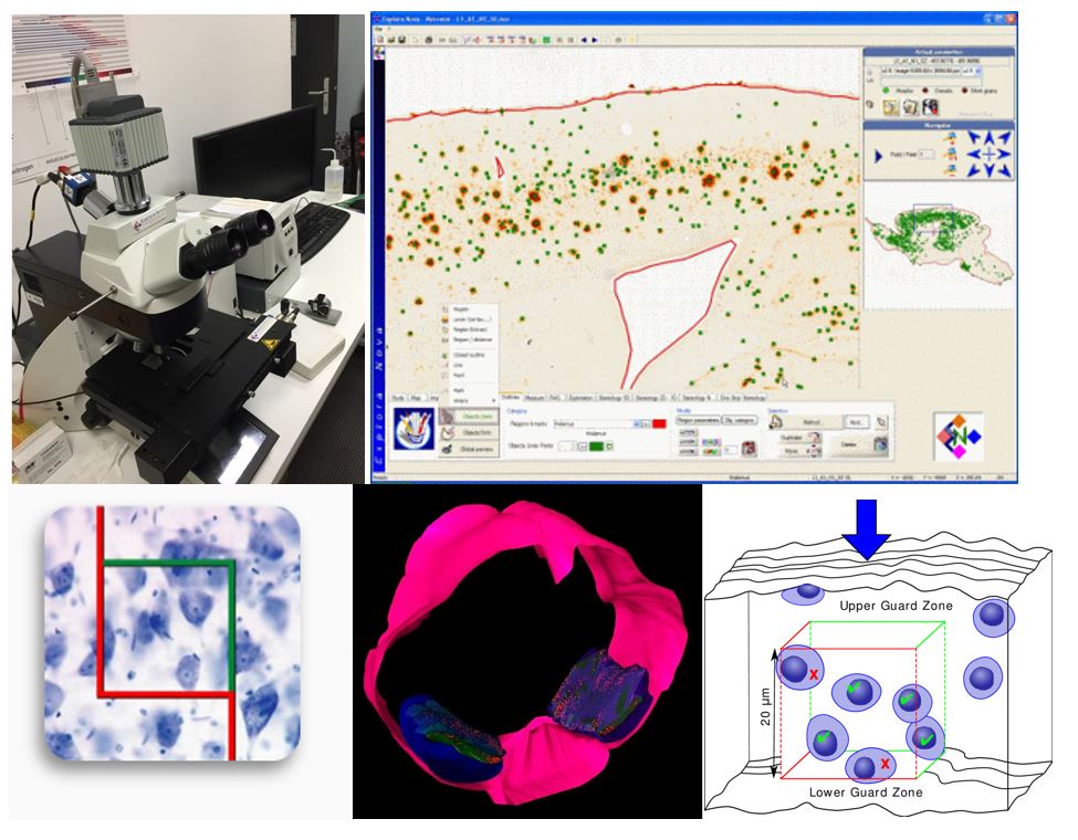
Mercator is a complete software for the quantification of histological sections. It allows you to define the contours of regions of interest at low magnification and to quantify high magnification cells. It allows to make morphological, colorimetric, densitometric evaluations, as well as stereological analysis. Mercator also eliminates the risk of double counting, and can generate high-resolution cards.
Imaris is a proprietary 3D / 4D reconstruction software, edited by Bitplane.
It makes it possible to visualize, images taken by light microscopy, by volume and surface reconstruction. The laboratory has its own license of Imaris (version 9.3), especially to analyze the data obtained with the microscope with sheet of light.
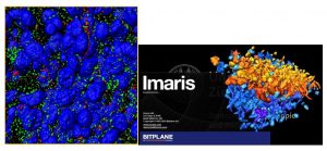
Plug-in :
contact: Eric Balado
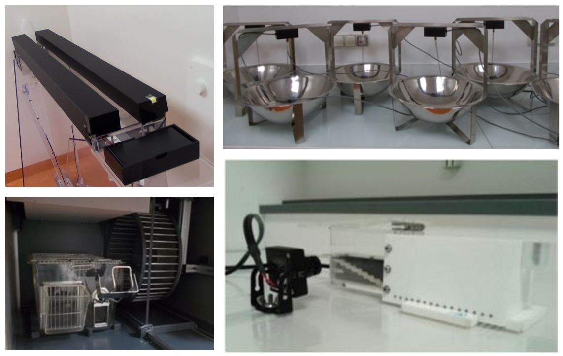
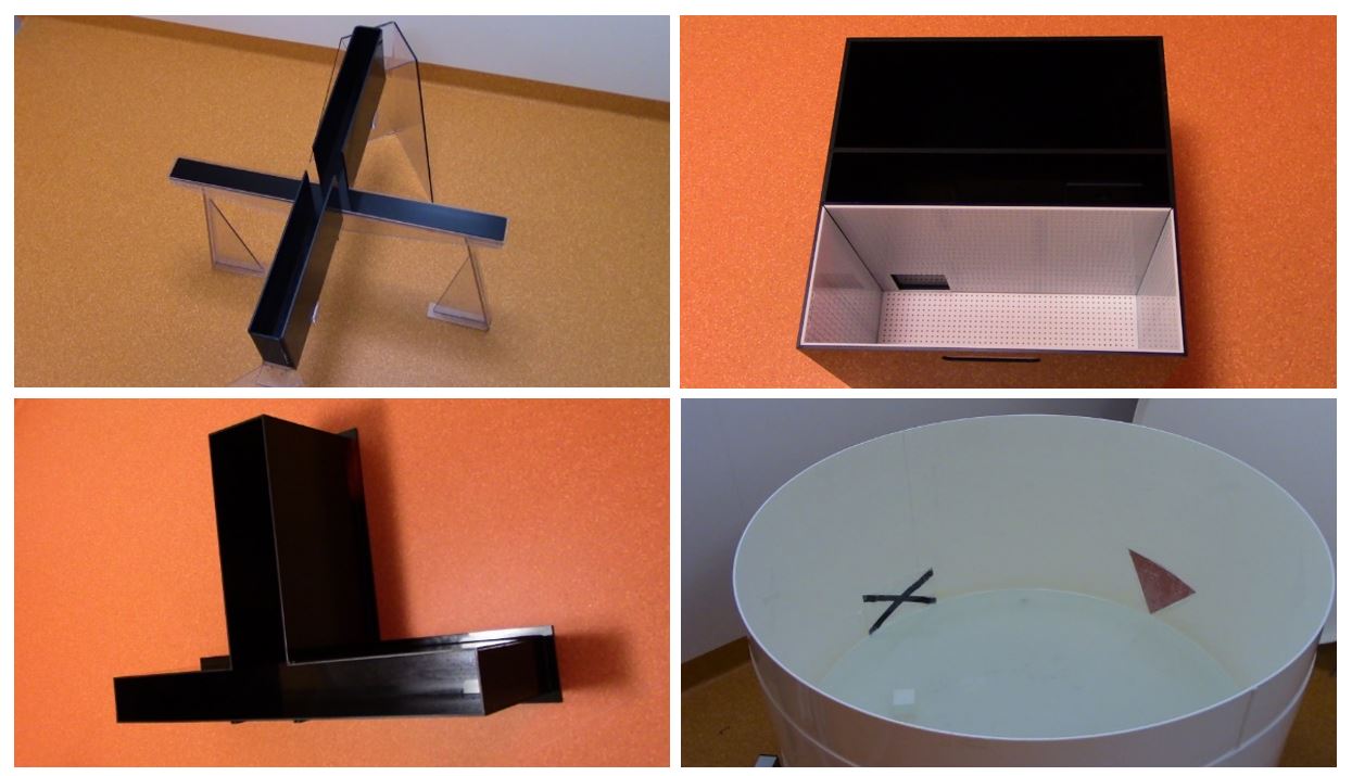
Addiction, comportement alimentaire et comportement cognitif :
contact : Marianne Benoit-Marand
The electrophysiology platform includes two in vivo recording stations and two brain slice recording stations.
In vivo electrophysiology equipment allows the extracellular recording of unit activity in neuronal action potentials in anesthetized rodents. The marking of neurons by neurobiotin during registration makes it possible to identify them a posteriori.
in vivo recording


The electrophysiology station includes a system for brain slices and an acquisition system for patch-clamp recording . This technique allows the study of membrane properties and neural synaptic responses
Ex vivo recording

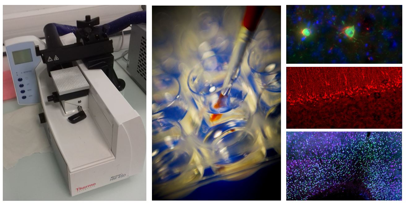
The histology platform brings together different cutting devices to adapt to a maximum of samples and applications: three microtomes, three vibratomes and two cryostats. This platform is used by all members of the three LNEC teams.
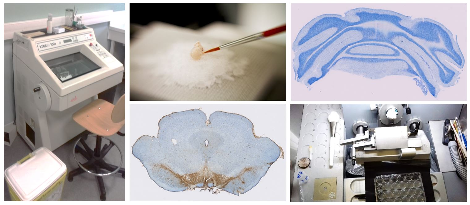 It makes it possible to obtain brain sections from rats or mice in order to carry out immunohistochemistry or in situ hybridization experiments.
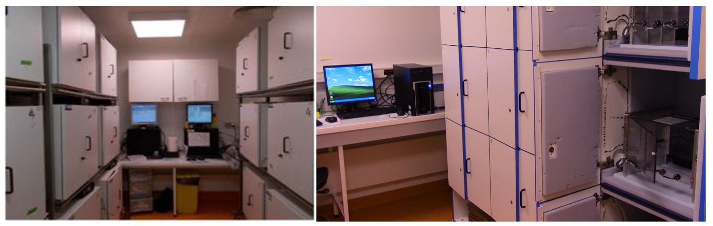
In 2013, freezers containing biological samples and antibodies were equipped with the SIRIUS Alarm System (JRI). Sirius is a software dedicated to monitoring the temperatures of a fixed installation.
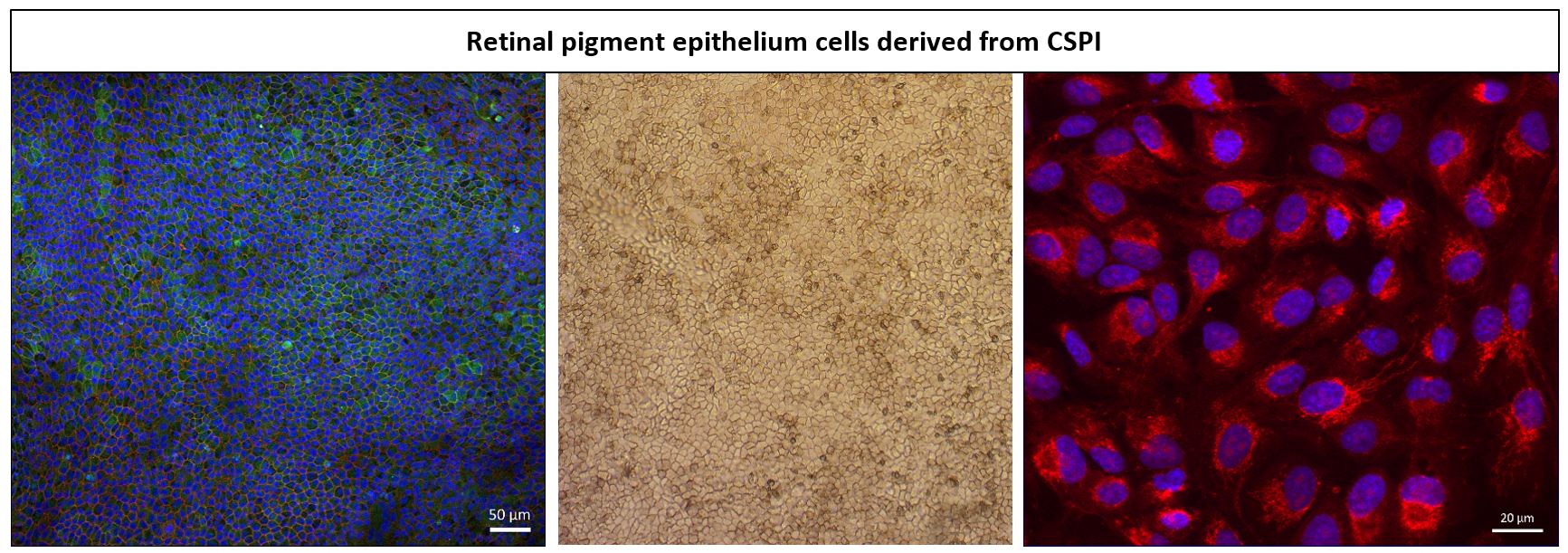
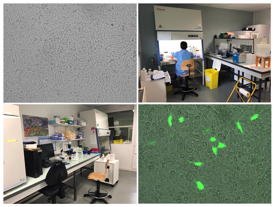
The laboratory has two cell culture pieces with HCB level C2 approval. To carry out the various cell culture projects, the rooms are equipped:
The platform is mainly used by members of Team 1 (A. Gaillard) for the development of protocols for differentiating stem cells into neural precursors, in the context of transplants in different animal models. This platform is also used in projects on glioblastoma and age-related macular degeneration.
The cell culture platform is also used by members of Team 3 (P.O. Fernagut) for the development and development of new studies requiring the transfection of murine cells with ShRNA and the primary culture of murine neurons.
contact : Virginie Lardeux
The HPLC platform is composed of an independent HPLC system with an electrochemical detector. The injection and chromatographic analysis of the samples are computer controlled. This platform is used to measure the concentrations of neurotransmitters and their metabolites obtained through microdialysis in vivo or in brain samples.
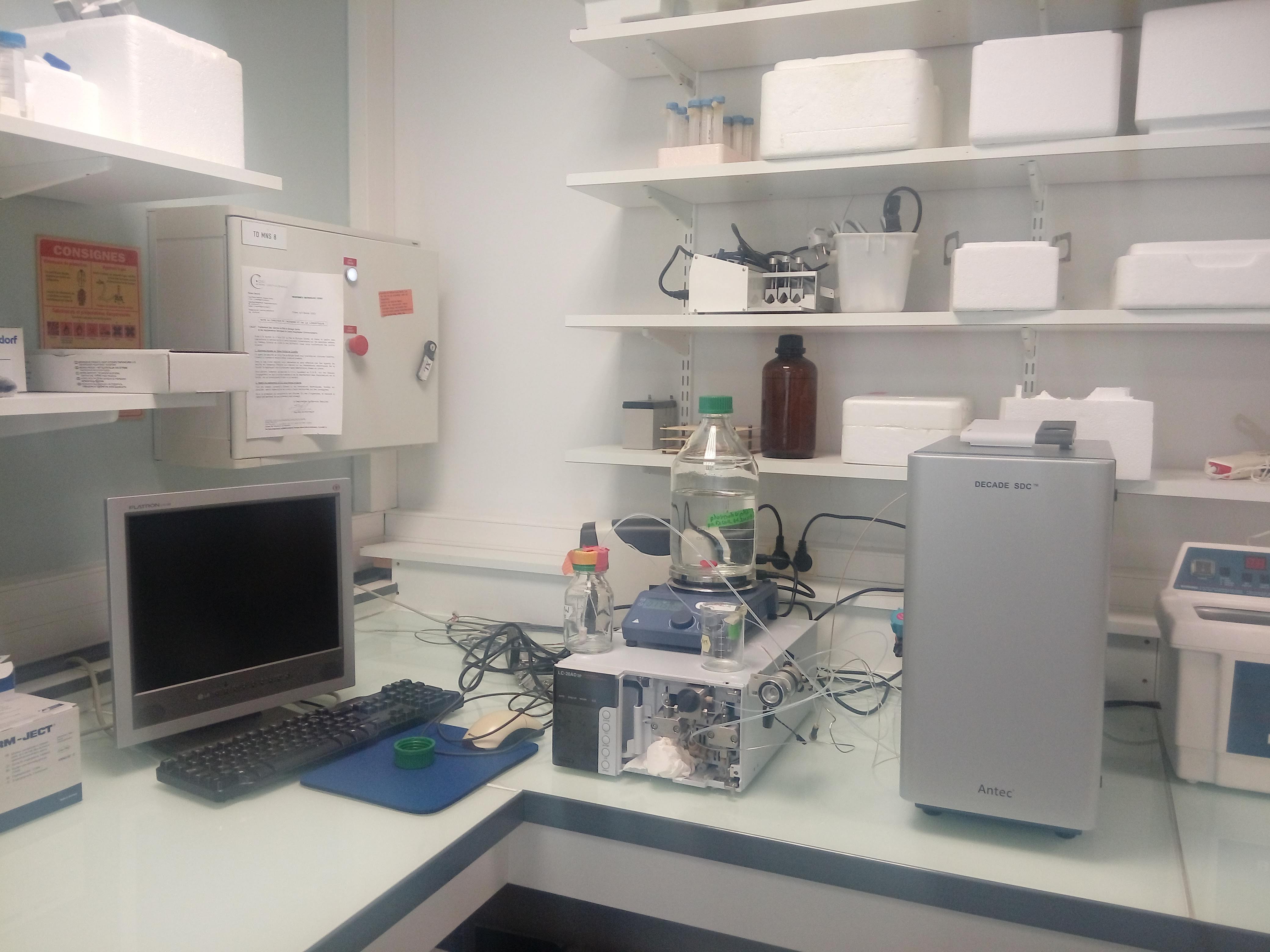
Système HPLC